News
News Release
Public Release of AI to Estimate Biological Sex from Fundus Images - Foundation for Better Understandings of Ocular Diseases Showing Sex Differences -
The Japanese Ophthalmological Society (JOS, President: OSHIKA Tetsuro, Tokyo, Japan) and the National Institute of Informatics (NII, Director-General: KUROHASHI Sadao, Tokyo, Japan) have developed and public-released an AI model to estimate an individual's sex from fundus images, using data collected by the Japan Ocular Imaging Registry (JOIR), a national ophthalmological database established with support from the Japanese Agency for Medical and Health Sciences (AMED). We expect that researchers will apply this AI model in the future research, and it helps elucidate the pathophysiology of diseases where the frequency of onset differs depending on biological sex.
Background
Deep learning has emerged as a significant breakthrough in machine learning and is widely used in artificial intelligence (AI), particularly in image recognition. Numerous reports suggest that the accuracy of the image recognition has surpassed that of humans. In 2017, NII established the Research Center for Medical Bigdata (RCMB) to develop medical assistance AI and created a database of medical image big data. NII also developed and operates an integrated cloud environment (cloud platform) equipped with high capacity data servers and GPU servers for machine learning calculation. Medical image analysis researchers across Japan, including Nagoya University, are connected to the cloud platform as part of NII's analysis team, and are collaborating to conduct research and development of various medical assistance AIs. In 2019, JOS established the General Incorporated Association Japan Ocular Imaging Registry (JOI Registry or JOIR) to collect various ophthalmic data from ophthalmology-related facilities nationwide, aiming to promote and support research and development of medical assistance AI. The collected ophthalmic images in the JOIR database are anonymized and sent to NII's cloud infrastructure to develop medical support AI using available computing resources on the cloud infrastructure.
In recent years, the development of AI using medical images has progressed rapidly, and it has become clear that it is possible not only to determine the presence or absence of a disease, but also to estimate the condition of the individual whose image was taken. It has become clear that AI can be used to estimate age, sex, smoking status, blood sugar level, etc. from fundus images taken of the retina, which is the light-sensing part of the eye, and the information obtained from this is used by ophthalmologists. It has the potential to be used in medical research targeting not only regional diseases but also various systemic diseases. However, in the research reported so far, the developed AI has not been made public, so it has not been possible to use it for other researches. To address this issue, JOS collaborated with NII to develop an AI that estimates a person's age based on fundus images, and in January 2023, the AI model (estimation method) has been made available to a wide range of researchers without any charge. As the second step, we have developed an AI that estimates biological sex from fundus images and are making it available to the public free of charge.
Outline
The model we developed used 131,031 fundus images of people between the ages of 17 and 94 with sex labels as training data, and performed deep learning with gender as the correct answer. During training, we used 16 popular deep learning models (DenseNet-121/169/201, Inception-V3, Inception-ResNet-V2, MobileNet, MobileNetV2, Xception, EfficientNet-B0/B1/B2/B3/B4/B5/B6/B7).
As a result, the accuracy with which the sex estimated from the fundus images of the validation data matched the actual gender was 92.0% (AUC 0.971) for the highest model, which is equivalent to the gender estimation models used for other races (Figure 1). Among these, we will publish two models: EfficientNet-B7, which was the most accurate, and MobileNet, which was lightweight and highly accurate.
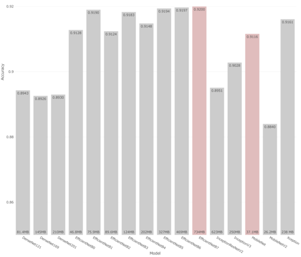 Figure 1 : Estimation accuracy of the sex estimation AI model developed this time
(bar basis: file size of model)
By utilizing these AI models, researchers may elucidate the pathophysiology of diseases whose onset frequency differs depending on biological sex, or supplement information when the sex information is missing in their research.
Public Release of the Model
The model can be downloaded from JOIR's webpage at the following URL:
http://www.joir.jp/data/index.html
Funding and Support
This study received support from AMED Clinical Research and ICT Infrastructure Development and Artificial Intelligence Implementation Research Project, "Establishment of Infrastructure for Image and Data Databases using ICT/Artificial Intelligence to achieve Next-Generation Ophthalmology" (Project Number: JP17lk1010024, Principal Investigator: OSHIKA Tetsuro), as well as "Research on Cloud Infrastructure and AI Image Analysis to Promote the Use of Medical Big Data" (Project Numbers: JP18lk1010028 and JP19lk1010036, Principal Investigator: AIDA Kento). The AI models were developed by Kensaku Mori (Director of NII RCMB and Professor at Nagoya University) and Masahiro Oda (Associate Professor at Nagoya University).
News Release: PDF
Links
SPECIAL
 NII Today No.101(EN)
 SINETStream Use Case: Mobile Animal Laboratory [Bio-Innovation Research Center, Tokushima Univ.]
 NII Today No.100(EN)
 Overview of NII 2023
 NII Today No.99(EN)
 Guidance of Informatics Program, SOKENDAI 23-24
 NII Today No.98(EN)
 The National Institute of Information Basic Principles of Respect for LGBTQ
 DAAD
DAAD